
▌X-ray Micro-Computed Tomography for Structural Analysis of All-Solid-State Battery at Pouch Cell Level
X 射線顯微電腦斷層掃描用於軟包級全固態電池之結構分析
Chen-Jui Huang, Jin An Sam Oh, Marta Vicencio, Tianchen Hu, Hedi Yang, James N. Burrow, Yen-Fang Song, Gung-Chian Yin, Pavel Shevchenko, Kamila M. Wiaderek, Bing Joe Hwang, and Ying Shirley Meng*
https://doi.org/10.1021/acsenergylett.5c00956
SEED Member: Bing Joe Hwang
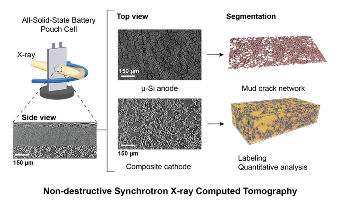
Non-destructive synchrotron X-ray computed tomography (sXCT) imaging of an ASSB pouch cell showing μ-Si anode, composite cathode, and 3D mud-crack network visualization.
Major Contributions
1.Establishment of synchrotron X-ray micro-computed tomography (sXCT) as a rapid, nondestructive, and high-resolution technique for visualizing and quantifying key microstructural features in all-solid-state pouch cells, including porosity, particle size distribution, contact loss, and delamination.
2.Demonstration that integrating sXCT-derived 3D models into multiphysics simulations enables predictive understanding of chemo-mechanical degradation, guiding the design of robust, high-performance solid-state batteries.
3.Advancement of imaging capabilities through computed laminography, multiscale operando imaging, and AI-assisted data processing, improving image quality, temporal resolution, and high-throughput analysis for industry-relevant pouch cell investigations.
主要貢獻
1.確立高效非破壞性檢測技術:確立了 同步輻射 X 射線顯微電腦斷層掃描(sXCT) 為一種快速、非破壞性且高解析度的技術。此技術能有效視覺化並量化全固態軟包電池中的關鍵微結構特徵,包含孔隙率(porosity)、粒徑分佈、接觸流失(contact loss)以及層間剝離(delamination)。
2.l結合模擬實現預測性設計:證實將 sXCT 建構的 3D 模型整合至 多物理場模擬(multiphysics simulations) 中,能實現對化學-機械衰退(chemo-mechanical degradation)機制的預測性理解,進而指導設計出更強韌且高性能的固態電池。
3.先進成像能力的拓展:透過引入電腦層析成像(computed laminography)、多尺度原位成像(multiscale operando imaging)及 AI 輔助數據處理,進一步提升了成像能力。這顯著改善了影像品質、時間解析度與高通量分析效率,使其更適用於具工業價值的軟包電池檢測。